
The Hallmarks of Tetralogy of Fallot
In compliance with the FTC guidelines, please assume the following about all links, posts, photos and other material on this website: (...)
The hallmarks of Tetralogy of Fallot
Tetralogy of Fallot is a form of a congenital heart defect that represents the combination of four heart defect symptoms that are present at birth. As a rare heart disease, it affects about 5 out of 10,000 children and it involves both genders equally. The classic physiological defect in the condition is the resulting abnormal blood flow through the heart owing to its structural defects. As a result, poor blood flow gets through the heart, depriving it with the proper nutrients and oxygen to carry out its normal function.
The hallmarks of Tetralogy of Fallot
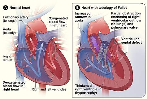
In Tetralogy of Fallot, not enough blood reaches the lungs in order to get sufficient oxygen to bring to the heart. As a result, poorly oxygenated blood reaches the heart. This is due to the four structural heart defects that are present in the disease that prevent the sufficient flow of oxygen throughout the body and in supplying the heart cells. These four structural defects of the heart are the hallmarks of Tetralogy of Fallot.

1. Pulmonary stenosis
This is a condition where there is a narrowing of the pulmonary valve that prevents the normal flow of the blood to get some oxygen supply from the lungs. With a narrow passage on the right ventricle, the oxygen poor blood from the right ventricles are unable to pass through in order to reach the lungs to get some oxygen supply to the blood.
2. Ventricular septal defect
The heart has a wall separating the two chambers located on either side of the heart which is called the septum. The septum plays an important role in preventing the mixture of the blood from both sides of the heart chambers. The presence of a ventricular septal defect causes a hole in the septum separating the lower left and right chambers called the ventricles. As a result, the non-oxygenated blood in the right ventricles mix with the oxygen rich blood in the left ventricles.
3. Right ventricular hypertrophy
This structural defect of the right ventricles of the heart produces thickened right ventricular muscles as a result of overloading the right ventricle in working harder in order to pump the blood through the narrowed right pulmonary valve (pulmonary stenosis).
4. Overriding aorta
The aorta is the part of the heart that carries the oxygen rich blood and is responsible for pumping the blood from the heart throughout the body. In the presence of the three other structural defects of the heart, the aorta has to work more than normal, thereby resulting in extremely fatigued and overworked aorta.
Symptoms of Tetralogy of Fallot
The common symptoms of the disease consist of cyanosis, a bluish looking skin color because of the lack of oxygen circulating in the body. Dizziness, fainting, and irregular heartbeat are common symptoms of the disease. Some may also experience seizures and delayed in growth and development due to the lack of oxygen circulating in the body. Those with Tetralogy of Fallot are also at higher risk of endocarditis.
Diagnosis and treatment
Proper diagnosis of Tretatology of Fallot is taken during pregnancy or when the child is born. Screening tests such as a prenatal examination can help diagnose the presence of birth defects. Ultrasound test can also identify abnormal heart development of the heart during pregnancy. A positive findings of heart abnormalities are usually confirmed by a fetal echocardiogram, which monitors the fetal heartbeat. Babies that are born with Tetratoly of Fallot usually manifest an episode of turning blue when feeding or crying. Common telltale signs and symptoms of the disease is a bluish looking skin and a whooshing sound called a heart murmur upon examination. Surgical intervention is the treatment for Tetralogy of Fallot after childbirth. The defective heart structures are replaced or widened in order to improve the flow of blood and oxygen through the heart.